
AEchocardiography (ECHO) is a non-invasive imaging test that uses ultrasound waves to create real-time images of the heart. It helps doctors see the heart’s structure, movement, and blood flow, providing vital information about how well the heart and its valves are working.
🔹 What ECHO Shows
An ECHO allows cardiologists to:
Visualize the size and shape of the heart
Assess heart pumping function (ejection fraction)
Detect leaking or narrowed valves
Identify congenital heart defects
Monitor fluid buildup around the heart (pericardial effusion)
Evaluate heart muscle strength and wall motion
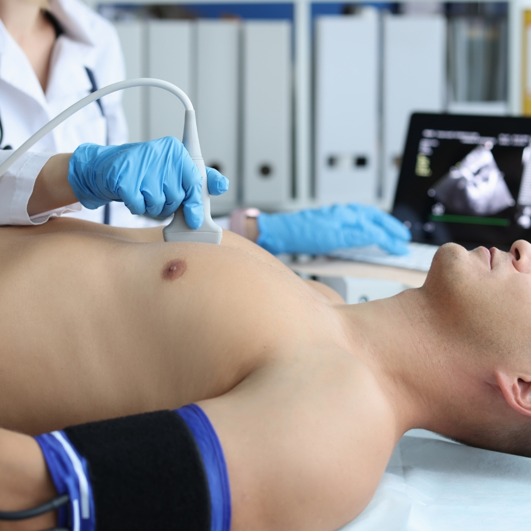
Echocardiography, often called ECHO, is a painless and non-invasive test that uses ultrasound waves to create moving images of the heart. It helps doctors evaluate the size, shape, and function of the heart and its chambers.
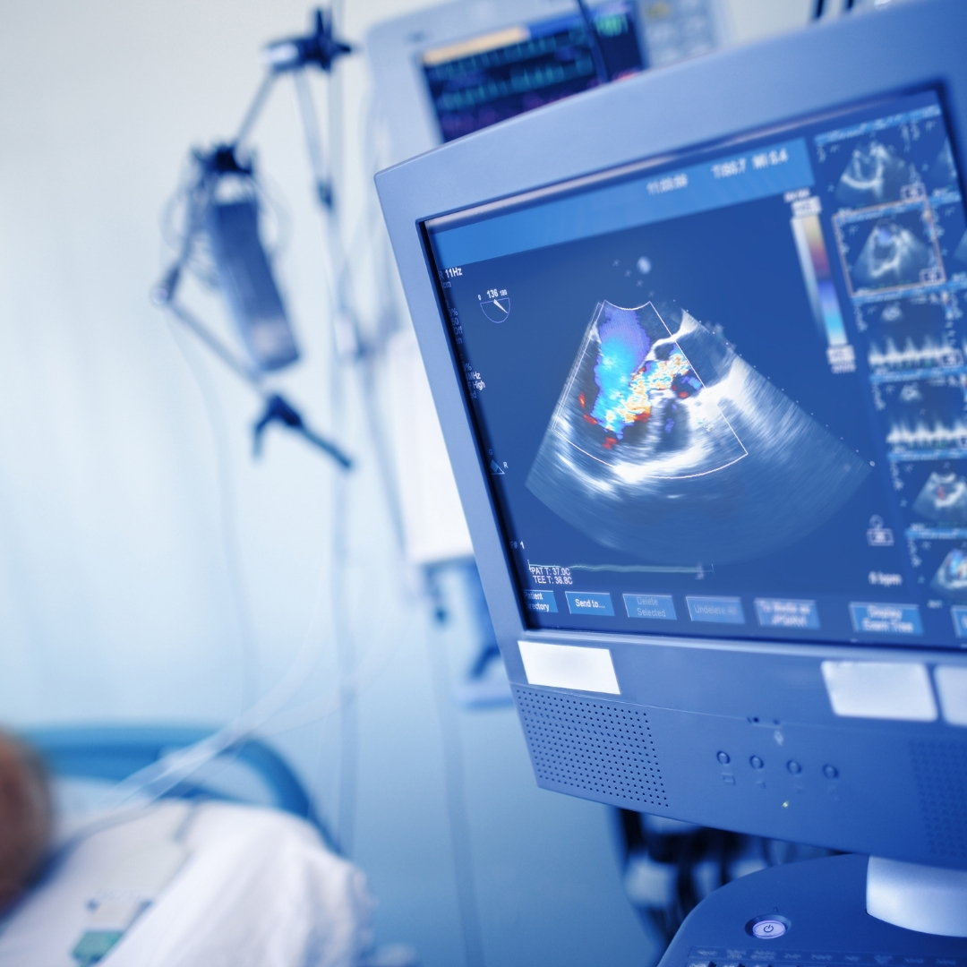
ECHO shows how well the heart muscles and valves are working and can detect problems like heart defects, valve disorders, and poor blood flow. It is an important diagnostic tool used in cardiology to monitor heart disease, congenital defects, or the effectiveness of treatment.
🔹Echocardiography helps doctors:
Evaluate the overall functioning of the heart.
Detect heart valve diseases and abnormal blood flow.
Identify congenital heart defects (present from birth).
Assess damage after a heart attack.
Monitor heart failure, cardiomyopathy, or infections of the heart valves
Check the effectiveness of ongoing treatment.
Raghuraj Hospital is a leading multi-speciality and cardiac care hospital located in Mansarovar, Jaipur. Known for its advanced facilities and compassionate healthcare, the hospital is dedicated to providing high-quality medical services under one roof.
C-26, Vande-Mataram Road, Opp. Iskcon Temple Road, Near Hotel Hyatt Regency, Krishna Sagar Colony, Mansarovar, Jaipur, Rajasthan 302020
© Raghuraj Hospital. All Rights Reserved.
Designed by CH DIGITAL SOLUTION COMPANY