
Arthroscopy is a modern, minimally invasive surgical procedure used to diagnose and treat problems inside a joint. It allows orthopedic surgeons to see the internal structure of joints using a specialized instrument called an arthroscope — a thin, flexible tube fitted with a camera and light source. This procedure provides a clear, magnified view of the joint’s interior, helping doctors identify and treat conditions without making large incisions.
Arthroscopy is commonly performed on major joints such as the knee, shoulder, hip, elbow, ankle, and wrist. It is particularly useful for patients suffering from joint pain, stiffness, swelling, or movement restriction that does not improve with medication or physiotherapy.
During the procedure, the surgeon makes a few small incisions (usually 0.5–1 cm) around the affected joint. The arthroscope is inserted through one incision, and miniature surgical instruments are introduced through others. The real-time video images are displayed on a monitor, allowing precise diagnosis and treatment of joint problems.
After arthroscopy, patients are usually discharged the same day or within 24 hours. Mild pain or swelling is normal and can be managed with medication and ice packs. A structured physiotherapy program is started soon after surgery to restore strength, flexibility, and joint movement. Most patients resume normal activities within a few weeks, depending on the joint treated and the extent of repair.
At Raghuraj Hospital, we specialize in advanced arthroscopic procedures performed by highly skilled orthopedic surgeons using the latest high-definition imaging and minimally invasive instruments. Our state-of-the-art operating theatres are equipped with modern arthroscopy towers and specialized tools for precise joint repair and reconstruction.
The Anterior Cruciate Ligament (ACL) is one of the key ligaments that help stabilize the knee joint. It connects the thigh bone (femur) to the shin bone (tibia) and plays a vital role in controlling knee movement, especially during twisting, turning, and jumping actions. An ACL injury is one of the most common sports-related knee injuries and can occur due to sudden stops, changes in direction, awkward landings, or direct impacts during sports or accidents.
A torn ACL causes pain, swelling, instability, and loss of knee strength, making it difficult to perform routine or athletic activities. While minor sprains can sometimes be treated with rest, physiotherapy, and bracing, a complete tear usually requires surgical reconstruction to restore knee stability and prevent further joint damage.
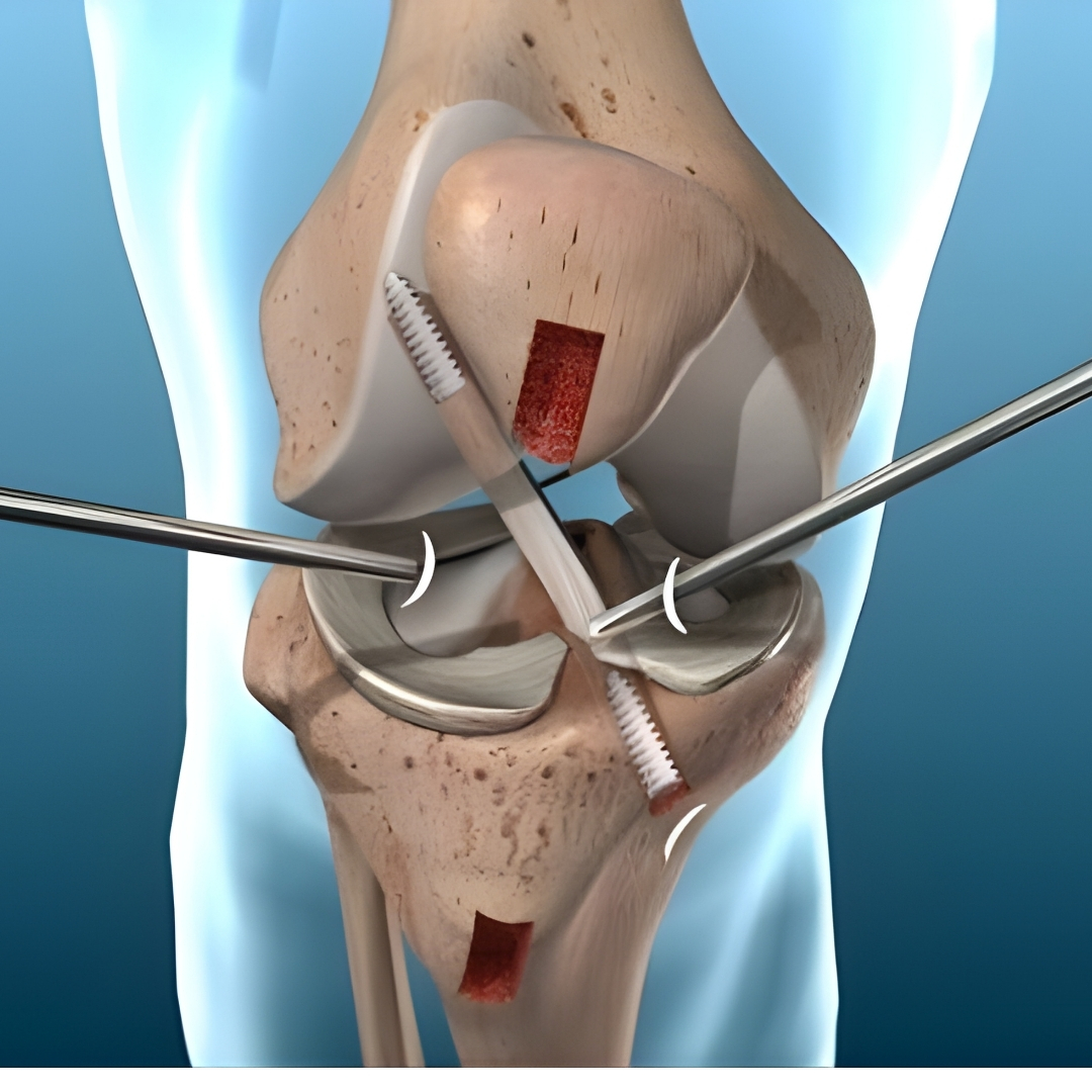
ACL Reconstruction Surgery involves replacing the torn ligament with a new one, known as a graft. This graft can be taken from the patient’s own body (autograft) — commonly from the hamstring or patellar tendon — or from a donor (allograft). The surgery is performed using arthroscopic (keyhole) techniques, which are minimally invasive and allow faster recovery.
During the procedure, small incisions are made around the knee to insert an arthroscope, which provides a clear, magnified view of the joint on a monitor. The surgeon removes the damaged ligament and creates tunnels in the femur and tibia to place the new graft securely with screws or fixation devices. The new ligament gradually integrates with the bone and functions like the natural ACL over time.
At Raghuraj Hospital, our orthopedic and sports injury specialists perform advanced arthroscopic ACL reconstruction using state-of-the-art equipment and modern graft fixation techniques. Our minimally invasive approach ensures minimal scarring, less postoperative pain, and quicker return to mobility.
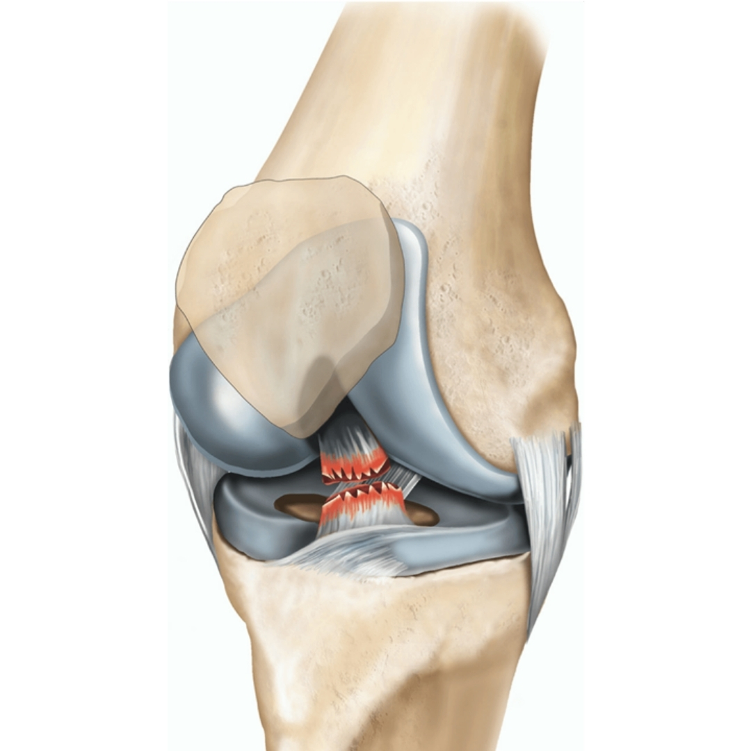
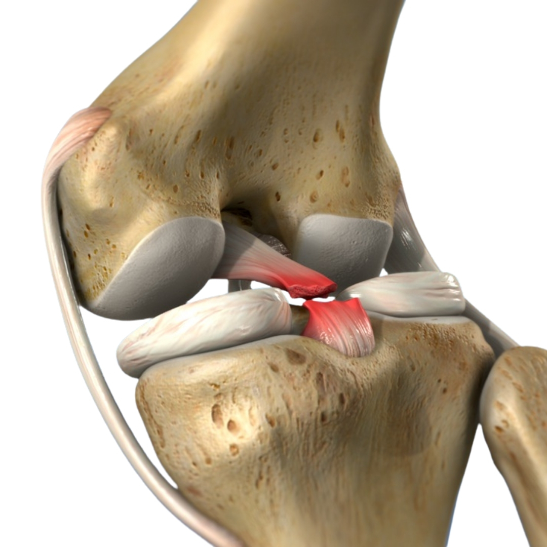
The Posterior Cruciate Ligament (PCL) is one of the four main ligaments that stabilize the knee joint. Located at the back of the knee, it connects the femur (thigh bone) to the tibia (shin bone) and prevents the tibia from moving too far backward. While PCL injuries are less common than ACL tears, they can significantly affect knee stability, strength, and movement when they occur.
A PCL injury often results from high-impact trauma, such as road accidents, sports collisions, or falls on a bent knee. Symptoms typically include pain, swelling, difficulty in bending the knee, instability, and a feeling of the knee "giving way" during movement. In many cases, PCL injuries occur along with damage to other ligaments or cartilage in the knee.
PCL tears are diagnosed through a detailed physical examination, specialized tests (such as the posterior drawer test), and imaging studies like X-rays and MRI scans. Early diagnosis helps determine whether conservative treatment or surgical reconstruction is needed.
Minor or partial PCL tears can often heal with rest, bracing, and physiotherapy, especially if the knee remains stable. However, complete or combined ligament tears, or cases where instability persists, require PCL reconstruction surgery to restore normal joint function.
PCL Reconstruction Surgery involves replacing the torn ligament with a graft, either taken from the patient’s own body (autograft) — such as the hamstring or quadriceps tendon — or from a donor (allograft). The procedure is performed using arthroscopic (keyhole) techniques, which are minimally invasive and allow precise visualization inside the knee joint.
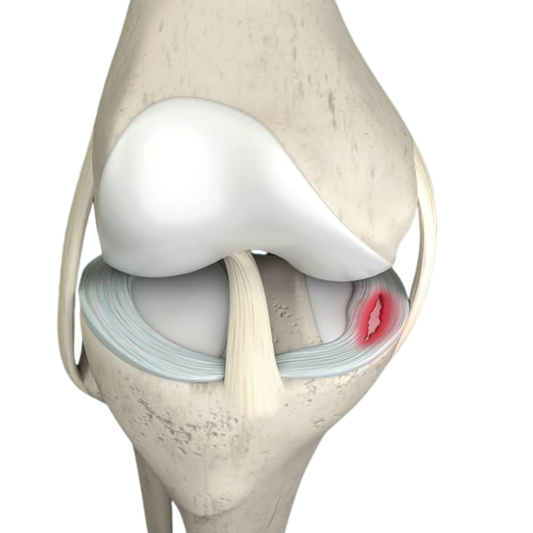
The meniscus is a C-shaped piece of cartilage located between the thigh bone (femur) and the shin bone (tibia) in the knee joint. Each knee has two menisci — the medial meniscus (inner side) and the lateral meniscus (outer side). These cartilage structures act as shock absorbers, distributing body weight across the knee, stabilizing the joint, and protecting it from wear and tear.
A meniscus tear is one of the most common knee injuries, particularly among athletes and individuals involved in activities that involve twisting, squatting, or sudden directional changes. It can also occur in older adults due to age-related cartilage degeneration.
A meniscus tear is diagnosed through a physical examination, along with imaging tests such as X-rays (to rule out bone injury) and MRI scans, which clearly show the extent and pattern of the cartilage tear.
After surgery, patients are advised to use crutches for a short period to avoid putting weight on the operated knee. Physiotherapy begins soon after to restore flexibility, muscle strength, and balance. Most individuals can return to regular activities within 4–6 weeks, and athletes can resume sports within 3–6 months after full recovery.
At Raghuraj Hospital, we offer advanced arthroscopic meniscus repair and reconstruction using state-of-the-art imaging and surgical technology. Our experienced orthopedic and sports injury specialists provide precise diagnosis, expert surgical care, and personalized rehabilitation programs to ensure complete recovery..
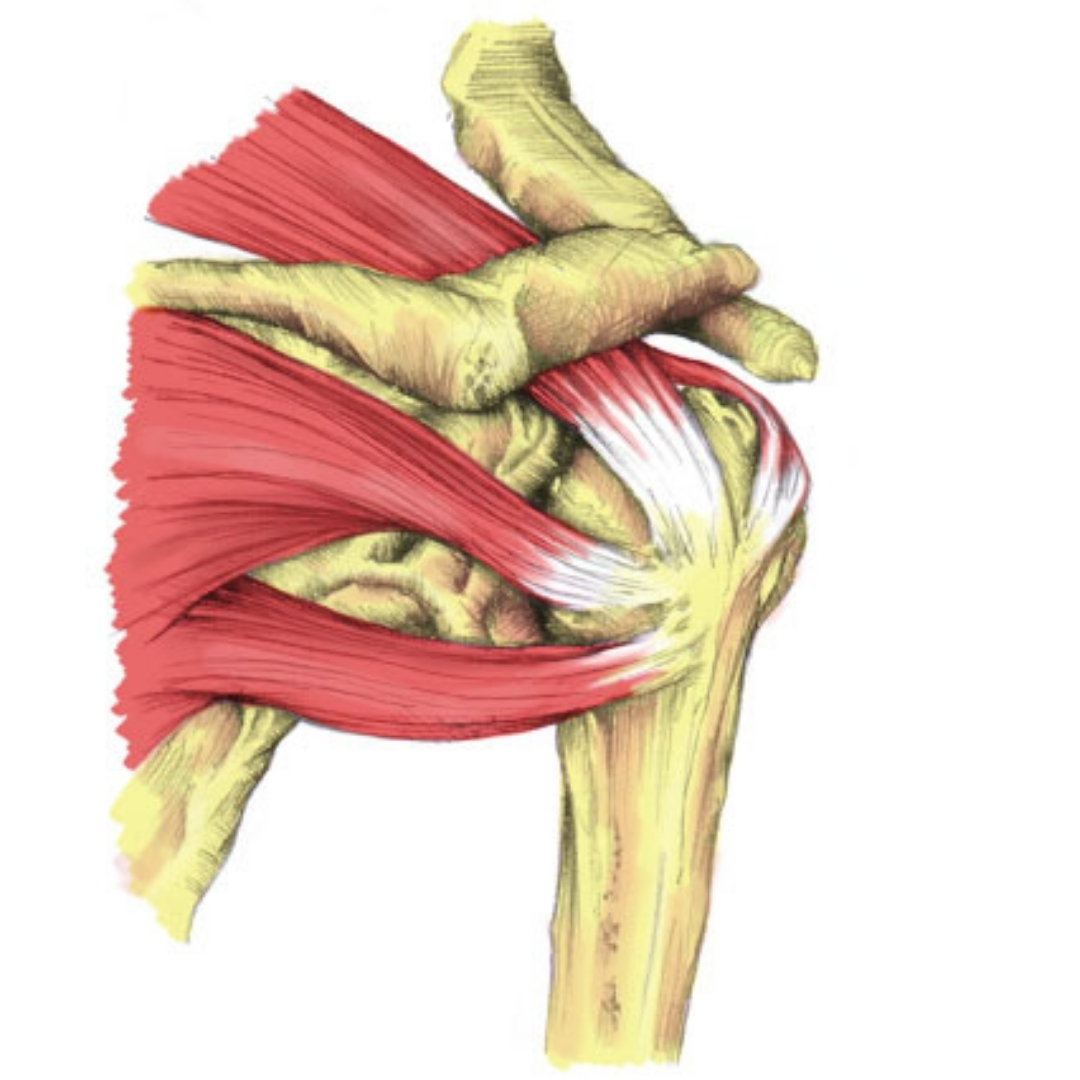
The rotator cuff is a group of four important muscles and their tendons that surround the shoulder joint — namely the supraspinatus, infraspinatus, teres minor, and subscapularis. Together, these structures stabilize the shoulder and allow a wide range of movements such as lifting, rotating, and reaching overhead.
A rotator cuff injury occurs when one or more of these tendons are inflamed, partially torn, or completely ruptured. This condition is one of the most common causes of shoulder pain and weakness, particularly in athletes, laborers, and older adults.
This minimally invasive technique uses a small camera (arthroscope) and fine instruments inserted through tiny incisions around the shoulder. The surgeon visualizes the torn tendon on a screen and carefully reattaches it to the bone using special sutures and anchors.
After surgery, the arm is supported in a sling for a few weeks to allow healing. Gradual physiotherapy helps restore shoulder movement, flexibility, and muscle strength. Patients typically regain full function within 3–6 months, depending on the extent of the tear and adherence to rehabilitation exercises.
At Raghuraj Hospital, our expert orthopedic surgeons specialize in arthroscopic shoulder surgery and use the latest technology to ensure precision and long-lasting results. Our dedicated physiotherapy and rehabilitation team designs customized recovery plans for each patient, ensuring a safe and complete return to normal life.
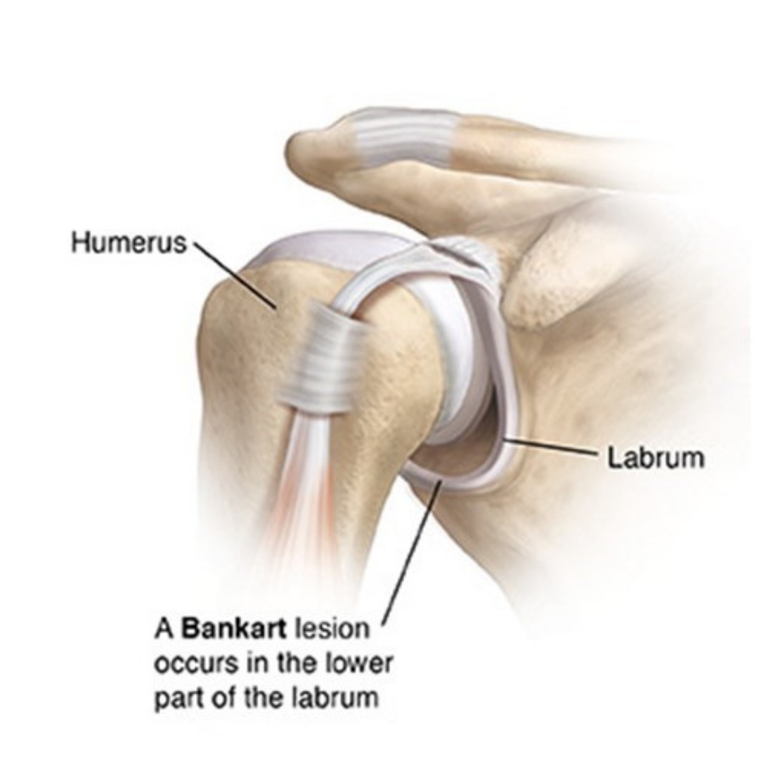
A Bankart repair is a specialized orthopedic procedure performed to restore stability to the shoulder joint after recurrent dislocations or chronic instability. The surgery specifically targets a Bankart lesion, which is a tear or detachment of the labrum — a ring of cartilage that lines the rim of the shoulder socket (glenoid).
The shoulder joint is a ball-and-socket structure that allows a wide range of motion. However, this mobility also makes it more prone to dislocations. When the shoulder dislocates, the head of the humerus (upper arm bone) moves out of the glenoid socket, often tearing the labrum and stretching the ligaments. If not properly repaired, this damage can cause repeated shoulder dislocations, pain, and weakness.
For mild instability, conservative treatment such as rest, physiotherapy, and strengthening exercises may be sufficient. However, for recurrent dislocations or labral tears, Bankart repair surgery becomes essential.
This minimally invasive surgery is performed using an arthroscope — a small camera inserted through tiny incisions in the shoulder. The surgeon visualizes the joint on a monitor and uses specialized instruments to:
At Raghuraj Hospital, we specialize in arthroscopic Bankart repair and shoulder stabilization procedures using state-of-the-art technology and high-quality suture anchors. Our expert orthopedic surgeons perform precision-based minimally invasive surgeries, ensuring excellent joint function and long-term results.
Raghuraj Hospital is a leading multi-speciality and cardiac care hospital located in Mansarovar, Jaipur. Known for its advanced facilities and compassionate healthcare, the hospital is dedicated to providing high-quality medical services under one roof.
C-26, Vande-Mataram Road, Opp. Iskcon Temple Road, Near Hotel Hyatt Regency, Krishna Sagar Colony, Mansarovar, Jaipur, Rajasthan 302020
© Raghuraj Hospital. All Rights Reserved.
Designed by CH DIGITAL SOLUTION COMPANY